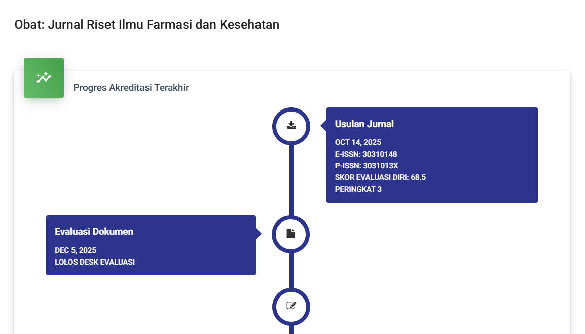
Evaluation of Some Neurotransmitters and Oxidative Stress by Manganese Chloride After Treatment with Lavender Ethanolic Extraction in Male Rats
DOI:
https://doi.org/10.61132/obat.v3i2.1113Keywords:
Serotonin, Lavender, Rats, Oxidative StressAbstract
This search intended to speculate the role of lavender ethanolic extract by reducing the toxicity of manganese in male rats. 32 white male Norwegian rats were divided into 4 equal groups. Group 1: as a control group left without treatments (only 1 ml of distilled water/ animal/ day). Group 2: were dosed manganese chloride at a concentration of 100 mg/ kg b.w. / day. Animals in Group 3 and Group 4 were dosed Manganese chloride at a concentration of 100 mg/kg b.w. then lavender ethanolic extract at a concentration of 200 and 400 mg/kg respectively, all by oral gavages and treated daily for six weeks. Then the next criteria were investigated: Neurotransmitters (dopamine and Ach (acetylcholine) in the mid brain) serotonin in serum. Oxidation indicators (glutathione and malondialdehyde in the brain and superoxide dismutase and catalase in serum). The results exhibited in both groups 3 and 4 there were a significant decrease in the concentrations of Ach accompanied by a significant increase in levels of dopamine and serotonin, also return levels of MDA (Malondialdehyde) to normal, as well as a significant increase in GSH (Glutathione) concentrations in rats mid brain. Moreover, the significant rising of SOD (Superoxide dismutase) and CAT (Catalase) levels in serum of rats in these groups indicated a noticeable improvement was achieved by lavender ethanolic extraction as compared to group 2. Conclusion, the antioxidant and antitoxic activity of lavender ethanolic extract promises in grate achievement in various health fields, including medicine, food industries and cosmetics.
Downloads
References
Andringa, K. K., Bajt, M. L., Jaeschke, H., & Bailey, S. M. (2008). Mitochondrial protein thiol modifications in acetaminophen hepatotoxicity: Effect on HMG-CoA synthase. Toxicology Letters, 177(3), 188–197. https://doi.org/[DOI]
Assessment, A. U. (2009). Experimental study on the effects of sodium selenite and N-acetylcysteine on oxidative damage of rat induced by manganese.
Barić, H., Đorđević, V., Cerovečki, I., & Trkulja, V. (2018). Complementary and alternative medicine treatments for generalized anxiety disorder: Systematic review and meta-analysis of randomized controlled trials. Advances in Therapy, 35, 261–288. https://doi.org/[DOI]
Blecharz-Klin, K., Piechal, A., Joniec-Maciejak, I., Pyrzanowska, J., & Widy-Tyszkiewicz, E. (2012). Effect of intranasal manganese administration on neurotransmission and spatial learning in rats. Toxicology and Applied Pharmacology, 265(1), 1–9. https://doi.org/[DOI]
Bouabid, S., Delaville, C., De Deurwaerdère, P., Lakhdar-Ghazal, N., & Benazzouz, A. (2014). Manganese-induced atypical parkinsonism is associated with altered basal ganglia activity and changes in tissue levels of monoamines in the rat. PLOS ONE, 9(6), e98952. https://doi.org/[DOI]
Bowman, A. B., Kwakye, G. F., Hernández, E. H., & Aschner, M. (2011). Role of manganese in neurodegenerative diseases. Journal of Trace Elements in Medicine and Biology, 25(4), 191–203. https://doi.org/[DOI]
Bryman, A., & Cramer, D. (2012). Quantitative data analysis with IBM SPSS 17, 18 & 19: A guide for social scientists. Routledge.
Chandel, M., & Jain, G. (2016). Manganese chloride induced hepatic and renal toxicity in Wistar rats. Toxicology International, 23(3), 212–220. https://doi.org/[DOI]
Chandel, M., & Jain, G. C. (2014). Toxic effects of transition metals on male reproductive system: A review. Journal of Environmental and Occupational Science, 3, 205. https://doi.org/[DOI]
Chis, I. C., Ungureanu, M. I., Marton, A., Simedrea, R., Muresan, A., Postescu, I. D., & Decea, N. (2009). Antioxidant effects of a grape seed extract in a rat model of diabetes mellitus. Diabetes and Vascular Disease Research, 6(3), 200–204. https://doi.org/[DOI]
Cordiano, R., Di Gioacchino, M., Mangifesta, R., Panzera, C., Gangemi, S., & Minciullo, P. L. (2023). Malondialdehyde as a potential oxidative stress marker for allergy-oriented diseases: An update. Molecules, 28(16), 5979. https://doi.org/[DOI]
Diederich, J., Brielmeier, M., Schwerdtle, T., & Michalke, B. (2012). Manganese and iron species in Sprague–Dawley rats exposed with MnCl₂∙4H₂O (IV). Microchemical Journal, 105, 115–123. https://doi.org/[DOI]
Dukhande, V. V., Malthankar-Phatak, G. H., Hugus, J. J., Daniels, C. K., & Lai, J. C. (2006). Manganese-induced neurotoxicity is differentially enhanced by glutathione depletion in astrocytoma and neuroblastoma cells. Neurochemical Research, 31, 1349–1357. https://doi.org/[DOI]
Farina, M., Avila, D. S., Da Rocha, J. B. T., & Aschner, M. (2013). Metals, oxidative stress and neurodegeneration: A focus on iron, manganese and mercury. Neurochemistry International, 62(5), 575–594. https://doi.org/[DOI]
Fitsanakis, V. A., Zhang, N., Anderson, J. G., Erikson, K. M., Avison, M. J., Gore, J. C., & Aschner, M. (2008). Measuring brain manganese and iron accumulation in rats following 14 weeks of low-dose manganese treatment using atomic absorption spectroscopy and magnetic resonance imaging. Toxicological Sciences, 103(1), 116–124. https://doi.org/[DOI]
Guilarte, T. R., et al. (2008). Impairment of nigrostriatal dopamine neurotransmission by manganese is mediated by presynaptic mechanisms: Implications to manganese‐induced parkinsonism. Journal of Neurochemistry, 107(5), 1236–1247. https://doi.org/[DOI]
Hancianu, M., Cioanca, O., Mihasan, M., & Hritcu, L. (2013). Neuroprotective effects of inhaled lavender oil on scopolamine-induced dementia via antioxidative activities in rats. Phytomedicine, 20(5), 446–452. https://doi.org/[DOI]
Hritcu, L., Cioanca, O., & Hancianu, M. (2012). Effects of lavender oil inhalation on improving scopolamine-induced spatial memory impairment in laboratory rats. Phytomedicine, 19(6), 529–534. https://doi.org/[DOI]
Jiang, Y., & Zheng, W. (2005). Cardiovascular toxicities upon manganese exposure. Cardiovascular Toxicology, 5, 345–354. https://doi.org/[DOI]
Kim, Y., et al. (2009). Effect of lavender oil on motor function and dopamine receptor expression in the olfactory bulb of mice. Journal of Ethnopharmacology, 125(1), 31–35. https://doi.org/[DOI]
López, V., et al. (2017). Exploring pharmacological mechanisms of lavender (Lavandula angustifolia) essential oil on central nervous system targets. Frontiers in Pharmacology, 8, 280. https://doi.org/[DOI]
Santamaria, A. (2008). Manganese exposure, essentiality & toxicity. Indian Journal of Medical Research, 128(4), 484–500.
Wang, J., et al. (2009). Expression changes of dopaminergic system-related genes in PC12 cells induced by manganese, silver, or copper nanoparticles. Neurotoxicology, 30(6), 926–933. https://doi.org/[DOI]
Zhang, S., Zhou, Z., & Fu, J. (2003). Effect of manganese chloride exposure on liver and brain mitochondria function in rats. Environmental Research, 93(2), 149–157. https://doi.org/[DOI]
Downloads
Published
How to Cite
Issue
Section
License
Copyright (c) 2025 OBAT: Jurnal Riset Ilmu Farmasi dan Kesehatan

This work is licensed under a Creative Commons Attribution-ShareAlike 4.0 International License.